Yeo2011_7Networks_MNI152_FreeSurfer 模板的正确打开方式
Forums
各位专家:
您好,我想用 Yeo2011_7Networks_MNI152_FreeSurfer 这个模板取出 ROI 来对我的一些数据做一些分析,可是这些模板用mricrond 打开以后的样子如附件截屏所示。不知道需要对结果图或者模板做怎样的转换才可以使用?多谢指教!
YUBING

各位专家:
您好,我想用 Yeo2011_7Networks_MNI152_FreeSurfer 这个模板取出 ROI 来对我的一些数据做一些分析,可是这些模板用mricrond 打开以后的样子如附件截屏所示。不知道需要对结果图或者模板做怎样的转换才可以使用?多谢指教!
YUBING
Hello!
I have some data gathering with the multi-band/Ts=2s(the filed_mapping_BOLD folder having 50 items), when I use the DPARSFA software for preprocessing, I was told not to do the "slice timing "step, but others said that I should do the "slice timing" in other software, and the data must be "slice timing". I was confused by those opposite view, what should I do then ? Can I use the data processing by the DPARSFA without "slice timing" to do the analyze?
Many thanks!
老师们好!
昨天发了一个英文版的问题,在论坛上没有找到,不知道是怎么回事,能帮忙看下吗?
先再把问题发一次,请教您两问题
1、跑DPARSFA运行到segment程序时就长时间没有响应了,也没有报错,处理结果文件夹中只有segment之前的文件,尝试多次都是出现附件图片中所示的问题。
2、DparsfA程序版本是否对Matlab兼容性有要求?对其他matlab toolbox版本是否有要求?我目前尝试了matlab2012和matlab2017版本。
期待您的回复!
谢谢啦!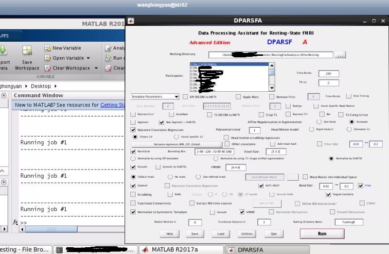
Hello!
I have two questions for help, thanks in advance~
1. when I used the DPARSFA on the matlab running my data, it worked well until the step of "segment", the matlab was not report "ERRORS", and the DPARSFA showed"RUN" state, I waited it for a long time (about 12 hours),but failed to find the processed data, the interface of the metlab just like the following picture:
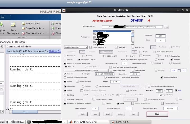
Hello, when I use FunImg as my starting directory, I get this error
"Too many .nii.gz files in each subject's directory, should only keep one 4D nii.gz file"
Both FunImg and T1Img were created in a previous run, so I don't understand why this error ?
appreciate any suggestions,
thank you.
Hi Dr. Yan,
What are the meaning of many variables of "cosub1_seg8.mat" in "T1ImgNewSegment" folder? Espeicially, does the value in "ll" means the total intra-cranial volume of a subjects?
If it is not, where can I find the brain sizes of all subjects?
Thanks
Hongru
严老师,
您好。最近学习R-fMRI network网站上的视频,帮助很大;论坛的讨论也给了我很多启发。但是最近在使用DAPBI的过程中也出现了一些问题。
问题描述:两组差异出现在了脑室。
1.自己按照论坛相关问题里面老师的回答,检查了标准化,操作,以及以往文献关于此疾病的处理过程。(见附件)
2.自己将DPABI结果与AFNI+FSL结果进行了对比,校正后结果不一致
自己的疑惑:
1,看到dpabi的Template文件夹中CSF的mask很小,我们用于regress的csf模板(选择的SPM先验模板)是这个模板吗,这个会造成脑室出现信号吗?
2.怎样确保自己处理数据的可靠性呢?个人逐个检查了标准化,按照老师在视频中说的标准,自己觉得配准没有出现不太好的结果。
3.如果处理的数据没有问题,怎么做一个合适的mask以去掉脑室进行统计呢,template中CSF的mask很小?